

In recent years, there has been a significant advancement in the field of Artificial Intelligence (AI) and Augmented Reality (AR). These technologies have become increasingly popular and have the potential to enhance virtual experiences in various fields such as gaming, education, healthcare, and...
A Neural Network Identifies Fungal Infections on Plants from a Single Image
136
26.07.2026
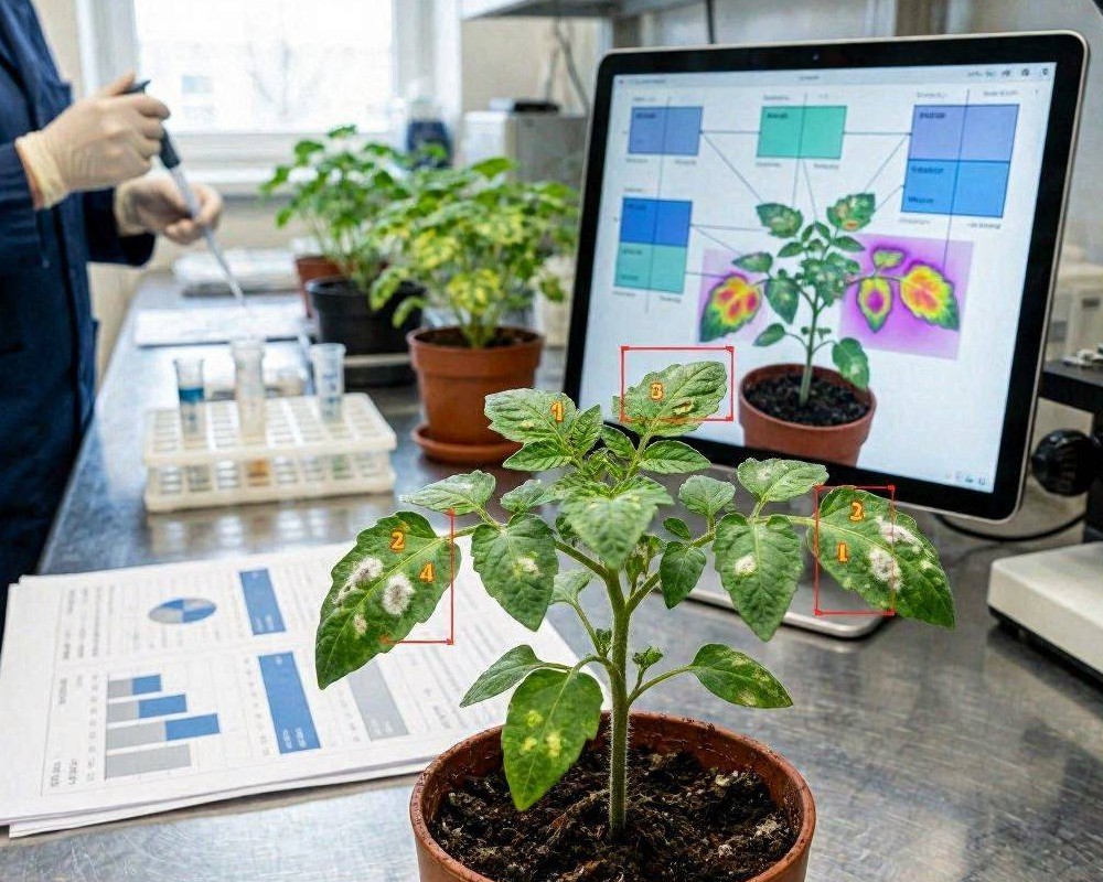
Fungal diseases remain one of the biggest threats to crop health, quality, and yield. Early detection is crucial: many infections progress quickly once symptoms appear. Traditionally, identification relies on expert visual inspection, laboratory tests, or time-consuming scouting. A growing body of applied AI research now shows a more practical alternative—using a neural network that can detect fungal infections from a single plant image.
Why single-image detection matters
In real farming conditions, images are often the only immediately available data. Weather changes, growth stages, and lighting differences make it hard to standardize observations. A model that works from one photo can reduce barriers by enabling fast triage: farmers, agronomists, and extension services can capture an image on-site and receive a likely diagnosis without waiting for lab results.
Single-image systems also support scalable workflows. Instead of sending samples for every suspicious case, operators can prioritize high-risk issues, target targeted treatments, and monitor disease spread across fields more efficiently.
How a neural network learns to recognize fungal infections
At the core of this approach is supervised machine learning. The model is trained on a large set of labeled plant images—examples where the disease type (or healthy condition) is known. During training, the network learns visual patterns associated with infection, such as lesion shape, color transitions, leaf texture changes, and distribution patterns across the plant surface.
Feature extraction with convolutional architectures
Most high-performing plant disease detectors use convolutional neural networks (CNNs) or modern transformer-based vision models. These architectures excel at extracting spatial features that correlate with disease manifestations. Even when symptoms vary between cultivars or environments, the model can capture consistent cues that humans might miss during fast field assessments.
Handling real-world image variability
For single-image inference to be reliable, the model must tolerate variability in:
- Illumination (sun glare, cloudy lighting, indoor capture)
- Camera quality and resolution
- Leaf angles and occlusions
- Background clutter (soil, stems, weeds)
- Growth stage differences
Training strategies like data augmentation (random crops, brightness changes, rotation, and blur simulation) help the network generalize. Some pipelines also use segmentation or region-focused pre-processing so the model emphasizes the leaf area rather than irrelevant background.
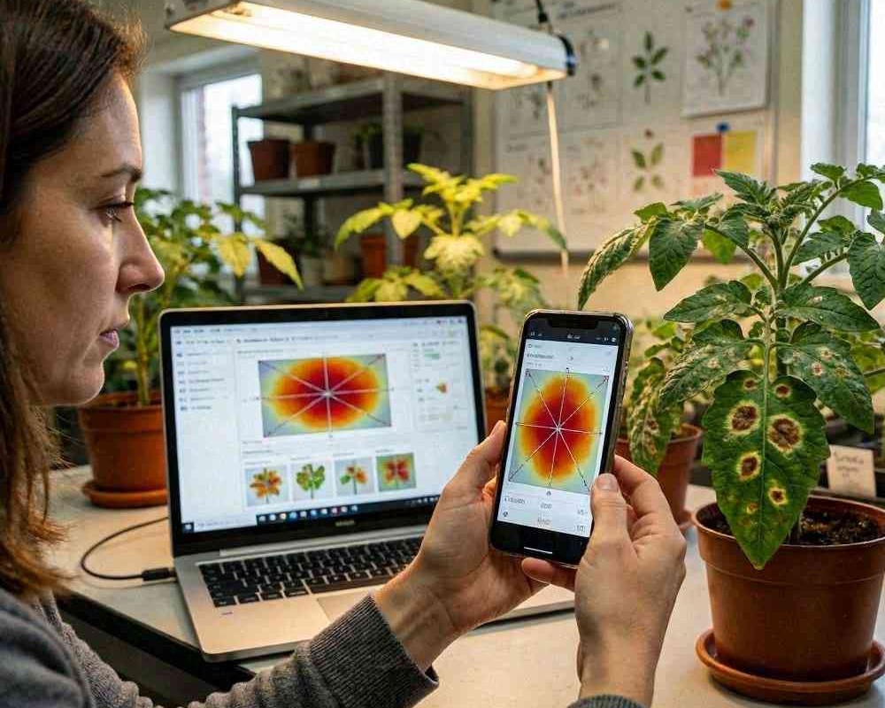
From photo to prediction: the inference workflow
Once trained, the workflow typically looks like this:
- Capture a clear image of the affected leaf or plant part, ideally filling most of the frame.
- Run the image through the neural network.
- Receive predicted disease classes (e.g., powdery mildew, leaf spot, rust) along with confidence scores.
- Optionally, export results into a field log or agronomic decision tool.
Confidence scores are especially important. Instead of presenting a single “answer,” a well-designed system provides uncertainty information so users can decide whether to trust the result, reshoot, or escalate to lab confirmation.
Interpreting confidence and avoiding false certainty
In agriculture, mistakes can be costly, so responsible AI design includes calibrated confidence. A prediction with low confidence may indicate that the symptoms are ambiguous, the image is too noisy, or the infection type is outside the model’s knowledge range. Including a “healthy/unknown” category, plus uncertainty-aware thresholds, helps reduce overconfident misdiagnoses.
Benefits for farmers, agronomists, and plant-health programs
A single-image fungal detection system can improve outcomes across multiple dimensions:
- Faster decisions: identify probable infections during scouting rather than waiting for lab timeframes.
- Reduced costs: prioritize samples for confirmation and avoid unnecessary chemical applications.
- Targeted management: align treatment strategies with the likely disease type and its recommended controls.
- Better monitoring: create a historical disease record for each field or block.
- Scalability: extend expertise using mobile capture and automated inference.
Limitations and what to do in practice
Despite strong performance, these systems are not a replacement for agronomic expertise. Symptoms can overlap across diseases, nutrient deficiencies, and pest damage. Environmental stress may produce leaf patterns that resemble fungal infections. Therefore, best practice is to treat model outputs as decision support, not absolute diagnosis.
To improve reliability:
- Use multiple photos from different angles when possible.
- Capture images across several plants in the same area.
- Compare results with local agronomic knowledge and disease calendars.
- Validate unusual or high-stakes cases with expert review or lab tests.
The future of image-based plant disease diagnostics
As datasets expand across regions, cultivars, and seasons, neural networks will become more robust and inclusive. Future systems may combine single-image diagnosis with additional signals—such as weather data, growth stage metadata, or multi-image sequences—to increase accuracy while keeping field workflows simple. For now, the ability to detect fungal infections from a single photo already represents a major step toward practical, AI-assisted plant protection.